
Introduction to Nerve Repair and Grafting
Peripheral nerves play a critical role in transmitting motor, sensory, and autonomic signals between the central nervous system—including the brain and spinal cord—and the rest of the body. There are different types of nerve: sensory nerves, which carry information from the periphery to the CNS; motor nerves, which transmit signals from the CNS to muscles; and mixed nerves, which contain both sensory and motor fibers and facilitate integrated functions. Nerves are composed of nerve fibers, which are the structural units responsible for carrying these signals.
When injured, damage to sensory nerves, motor nerves, or mixed nerves can result in lost function such as weakness, inability to move, sensory loss, chronic pain, or functional disability. This affects life profoundly. Unlike some tissues, nerves have a limited capacity to regenerate, and recovery depends greatly on timely and appropriate intervention. Nerve repair and grafting are the backbones in modern reconstructive surgery aimed to restore function after lost function due to injury. Nerve repair ensures continuity of the nerve, promotes axonal regeneration, and ultimately helps recover its function.
Direct nerve repair is possible when the gap between injured ends is minimal, while grafting is necessary for larger defects where tensionless repair cannot be achieved.
Over the past decades, advances in microsurgical techniques, bioengineered grafts, and adjunctive therapies have significantly improved outcomes. Understanding the principles, indications, and evolving methods of nerve repair is essential for surgeons and clinicians managing peripheral nerve injuries.
Historical Evolution of Nerve Repair
The history of nerve repair dates back to ancient civilizations, where early physicians attempted primitive suturing of severed nerves with limited understanding. In the 19th century, experimental studies demonstrated the potential for nerve regeneration, laying the foundation for modern nerve repair and joining. The introduction of the microscope in the mid-20th century brought in a very important tool in nerve repair. It made precise microsurgical repairs possible.
The advent of high quality Micro Needle Holders, Micro scissors, and Micro Forceps boosted microsurgery to a great extent.
Pioneering work on autologous nerve grafts [meaning using nerves taken from the patient’s own body] further advanced functional recovery in cases of nerve gaps.
More recently, bioengineered conduits, allografts [where a nerve is harvested from a donor], and adjunctive therapies have transformed outcomes, making nerve reconstruction an evolving specialty.
Anatomy and Physiology of Peripheral Nerves
Peripheral nerves are nerves that connect the central nervous system, including the spinal cord, to muscles, skin, and organs. They facilitate motor, sensory, and autonomic nervous system functions as part of the peripheral nervous system. Peripheral nerves include both spinal nerves, which emerge from the spinal cord, and cranial nerves, which connect directly to the brain. Cranial nerves play key roles in sensory and motor functions of the head and neck, and are distinct from spinal nerves.
Structurally, each nerve consists of bundles of axons, called fascicles, encased by connective tissue layers: the endoneurium surrounds individual axons [an axon is the long, slender projection of a single nerve cell (neuron) that conducts electrical impulses and serves as its functional unit for communication], the perineurium encloses fascicles, and the epineurium provides overall protection. A neuron consists of a cell body, dendrites, and an axon; the cell body contains the nucleus and is essential for the neuron's function. Nerve cells transmit information throughout the nervous system.
Axons are either myelinated, allowing rapid signal conduction, or unmyelinated, conducting slower impulses. The myelin sheath, formed by Schwann cells in the peripheral nervous system, speeds up electrical signal conduction along the axon. Schwann cells play a vital role in myelination and nerve regeneration after injury. Blood vessels within the epineurium ensure metabolic support and maintain blood flow, which is crucial for nerve health, while the perineurium maintains a protective barrier against mechanical and chemical insults.
Peripheral nerves carry signals as electrical impulses between the central nervous system and the body. Sensory nerves, as part of the peripheral nervous system, transmit sensory information such as touch, pain, and temperature from receptors to the CNS. Autonomic nerves regulate involuntary functions and control internal organs like the heart, lungs, and digestive system, maintaining homeostasis.
Understanding the three-dimensional organization, blood supply, and conduction physiology of peripheral nerves is essential for planning surgical repair and grafting. Nerve conduction studies are important diagnostic tools for assessing nerve function and identifying types of nerve injury. Knowledge of nerve branching helps surgeons optimize repair techniques, and enhance functional recovery after injury.
How Nerve Injuries Occur
Peripheral nerves can be damaged through a variety of mechanisms, each influencing the extent of functional loss and potential for recovery.
-
Traumatic injuries, such as lacerations, crush injuries, stretch injuries, or stretch, are the most common causes of peripheral nerve injury, often resulting from accidents, falls, or surgical mishaps. In some cases, a torn nerve may require surgical intervention to restore function.
-
Compression injuries, whether acute or chronic, can lead to ischemia and demyelination.
Nerve injuries are classified based on severity:
-
Neuropraxia involves temporary conduction block without axonal loss.
-
Axonotmesis includes axonal disruption with preserved connective tissue, allowing potential regeneration.
-
Neurotmesis represents complete nerve transection with loss of continuity. These cases require a surgical repair, especially when the injured nerve is severely affected.
-
Metabolic, infectious, and inflammatory conditions, such as diabetes or autoimmune neuropathies, can also impair nerve function. Peripheral neuropathy is commonly associated with these chronic conditions and may result from long-term complications of systemic diseases.
Nerve injury can result in damaged nerve tissue and nerve damage, which may present as loss of function or abnormal sensations. Symptoms of nerve damage can include numbness, tingling, weakness, and sharp pain, depending on the type and location of the injured nerve.
Understanding these mechanisms is crucial for determining the timing and type of intervention, guiding repair strategies, and predicting outcomes, as each mechanism uniquely affects axonal survival, regeneration potential, and ultimate functional recovery.
Principles of Nerve Healing and Regeneration
Peripheral nerve healing is a slow biological process aimed at restoring structural continuity and functional capacity.
Following injury:
-
Wallerian degeneration occurs distal to the site, where axons and myelin break down, clearing the path for regeneration.
-
Damaged tissue is carefully removed from the ends of a severed nerve before repair to facilitate optimal healing.
-
Schwann cells proliferate and form Büngner bands, providing guidance for regenerating axonal sprouts.
-
Axonal regeneration occurs at a rate of approximately 1 to 3 mm per day, influenced by patient age, injury type, and local microenvironment. Smaller nerves may regenerate differently or at different rates compared to larger nerves.
-
Tension-free alignment of nerve ends is critical for successful repair, as excessive tension impairs vascularity and axonal growth.
-
Neurotrophic factors, extracellular matrix proteins, and adequate blood supply support regeneration and remyelination.
-
Maintaining a healthy nerve and overall well-being is important for optimal recovery, as functional recovery also depends on timely repair, avoidance of scar formation, and early rehabilitation to prevent muscle atrophy.
Understanding these principles underpins surgical strategies and guides clinical decision-making, enabling surgeons to maximize the likelihood of meaningful sensory and motor restoration and to restore function.
Factors Influencing Success of Nerve Repair
The success of nerve repair is influenced by several factors, including the selection of appropriate nerve tissue and donor nerve for grafting procedures.
-
Timing of intervention is critical; early repair generally yields better outcomes.
-
Patient age, overall health, and comorbidities, such as diabetes, affect regenerative capacity.
-
The type and severity of injury, gap length, and degree of axonal loss influence matter and decide repair strategy.
-
Surgical technique is based on tension-free alignment, precise fascicular matching, and adequate vascularity.
-
When nerve grafts are required, choosing a suitable donor nerve and ensuring the quality of the harvested nerve tissue are essential for optimal repair.
-
Postoperative care, physiotherapy, and prevention of scar formation are crucial for functional recovery.
-
Additionally prevention of infection and ischemia is a must. A clean wound and good vascularity can promote regeneration, and presence of infection and poor blood supply will hinder regeneration.
-
This highlights the need for careful patient selection and meticulous surgical planning.
When to do Nerve Repair and Grafting: Indications
Nerve repair and grafting are indicated when functional recovery is unlikely without surgical intervention.
-
Direct repair is preferred in clean, sharp lacerations where nerve ends can be approximated without tension, typically seen in surgical or penetrating injuries.
-
Grafting becomes necessary when there is a segmental defect, such as after avulsion, crush, or delayed repair, where end-to-end approximation is not feasible.
-
Traumatic injuries with loss of continuity, iatrogenic nerve transections, and resection of neuromas or tumors often require grafting to bridge the gap.
-
Timing is very important, early intervention is favored, but delayed reconstruction may still offer benefits if target organs remain viable.
-
Progressive neurological deficits, persistent pain, and functional disability are strong indications for operative repair.
-
Careful patient evaluation, imaging, and intraoperative exploration guide the decision between direct repair, grafting, or alternative strategies, with the ultimate goal of restoring meaningful sensory and motor function.
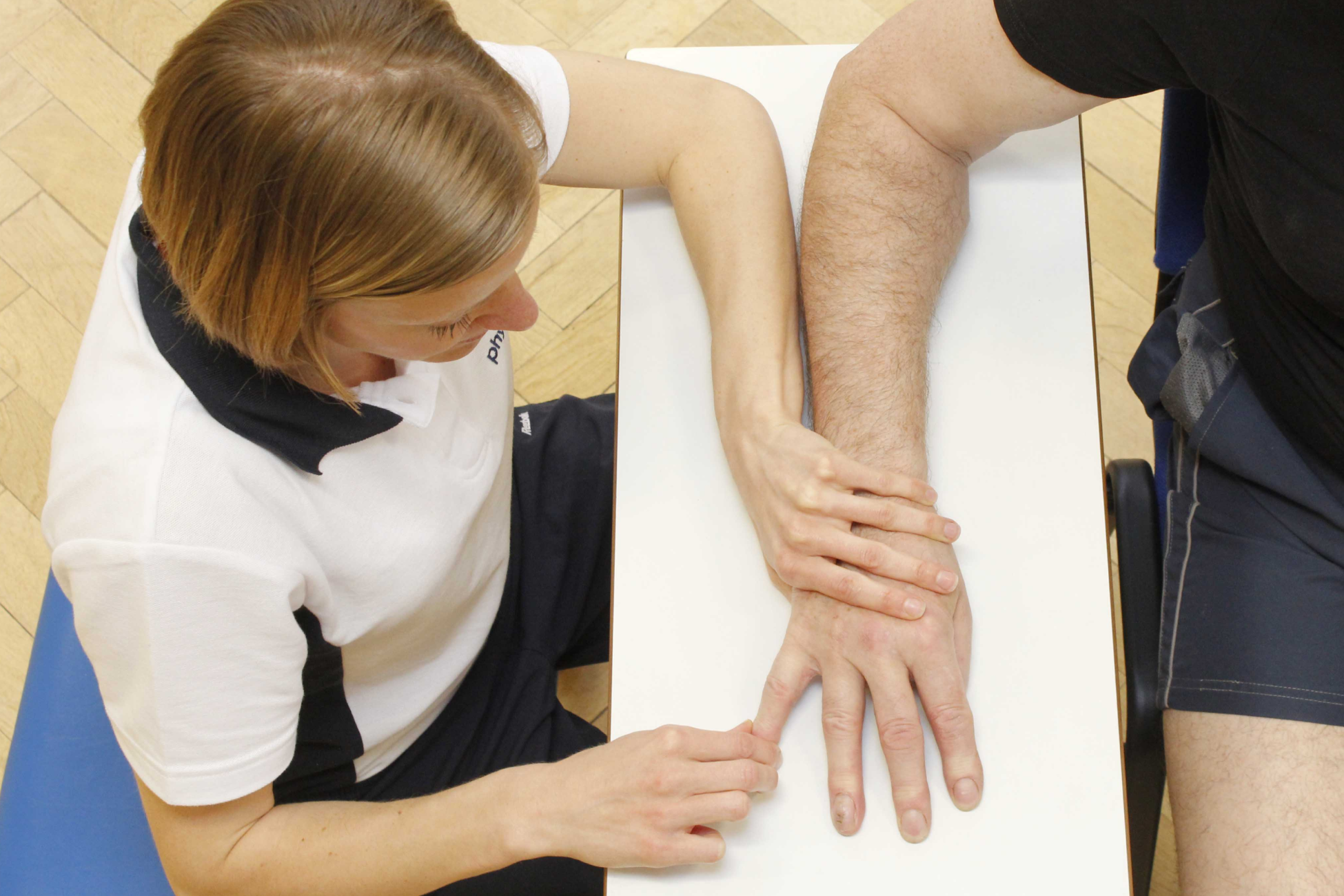
Techniques of Direct Nerve Repair
-
Direct nerve repair is called neurorrhaphy.
-
It is used when nerve ends can be approximated without tension.
-
The fundamental principle is precise alignment of the epineurium or fascicles to allow regenerating axons to cross the repair site effectively.
-
Epineurial repair, the most common technique, involves suturing the outer epineurium of the nerve ends with fine microsutures under magnification.
-
Microsurgery instruments like Micro Needle Holders, Micro scissors, and Micro Forceps are used.
-
Although expensive, such microsurgery instruments are also manufactured in countries like India. So high quality instruments are available at far more competitive prices compared to European manufacturers.
-
Indian startup Shira Medtech is one of the innovative manufacturers of Microsurgery instruments and has patented many advanced microsurgery instruments.
-
Fascicular repair, though technically more demanding, aligns individual fascicles for improved specificity in selected cases.
-
For smaller nerves, different techniques or specialized tools may be required, as standard suturing can be challenging due to their delicate structure.
-
Tension at the repair site must be avoided, as it impairs vascularity and axonal regeneration.
-
Microsurgical instruments, high-magnification optics, and atraumatic handling are critical for success.
-
Fibrin glue may be used to reinforce sutures or minimize manipulation.
-
Post-repair, the nerve is positioned to reduce stretch or compression.
-
If properly executed, direct repair offers the best chance for functional recovery.
-
However, when gaps are present, nerve grafting or conduits become necessary.
Nerve Grafting: Types, Sources, and Methods
-
Nerve grafting is required when there is a gap between severed nerve ends and it prevents tension-free repair.
-
The goal is to provide a biological scaffold through which regenerating axons can grow toward target tissues.
-
Autologous nerve grafts, harvested from donor sites such as the sural, medial antebrachial cutaneous, or great auricular nerves, remain the gold standard due to their biocompatibility and presence of Schwann cells. Selection of a donor nerve is based on factors such as function, location, and the availability of healthy nerve tissue, ensuring minimal loss of sensation or function in non-critical areas.
-
Drawbacks of autologous grafts are: donor site injury and possible loss of function; and limited availability.
-
Allografts, obtained from cadaveric donors, offer an alternative when long grafts are required
-
But allografts often need immunosuppression.
-
Synthetic conduits and bioengineered grafts are increasingly used, particularly for small gaps, and may incorporate growth factors or extracellular matrix proteins to enhance regeneration.
-
Methods of grafting include cable grafts for larger nerves, interfascicular grafts for selective fascicle alignment, and vascularized grafts for compromised beds. The structure of these grafts is designed to support the organization and regeneration of nerve fibers, which is critical for successful nerve repair.
-
The choice depends on defect length, nerve type (sensory, motor, or mixed), and clinical context. Different types of nerve may require tailored grafting approaches.
-
Common donor sites include the sural nerve, but the upper extremity is also frequently used for nerve grafting procedures, depending on the location and type of nerve injury.
Advances in Nerve Conduits and Bioengineered Grafts
Recent innovations in nerve repair have focused on developing alternatives to traditional autologous grafts, aiming to overcome limitations of donor site morbidity and limited graft length. Nerve conduits made of collagen, polyglycolic acid, or silicone provide a protective channel that guides axonal growth across gaps, particularly useful in defects up to 3 cm. Bioengineered grafts are being designed to mimic the natural extracellular matrix and may be seeded with various cell types involved in nerve regeneration, including nerve cells and Schwann cells, as well as stem cells or growth factors to accelerate regeneration. Advances in nanotechnology have enabled the incorporation of conductive polymers and electrical stimulation systems within conduits, further enhancing axonal guidance. The integration of electrical stimulation is particularly relevant at the neuromuscular junction, a specialized synapse where motor neuron terminals communicate with muscle fibers, playing a crucial role in motor control and nerve-muscle repair. Biodegradable scaffolds that degrade as regeneration progresses are also being researched.
Although these technologies show great promise in experimental and early clinical settings, long-term data on functional outcomes are still evolving. Yet, bioengineered grafts represent the cutting edge in peripheral nerve reconstruction.
Research and Experimental Approaches
Research in peripheral nerve repair is rapidly evolving, exploring strategies that go beyond traditional surgical techniques.
-
Stem cell therapy shows promise, with mesenchymal and induced pluripotent stem cells differentiating into Schwann-like cells that enhance regeneration. Various cell types, including nerve cells, are being studied for their roles in neural repair and regeneration.
-
Growth factors such as nerve growth factor (NGF) and brain-derived neurotrophic factor (BDNF) are being incorporated into conduits or delivered via controlled-release systems to stimulate axonal growth.
-
Tissue engineering approaches combine biomaterials with cellular and molecular therapies to create living scaffolds.
-
Electrical stimulation is another experimental modality shown to accelerate axonal sprouting and improve target reinnervation.
-
Gene therapy techniques are being investigated to upregulate regenerative pathways locally.
-
Advances in 3D printing allow the customization of nerve conduits that replicate natural fascicular architecture.
Although most of these strategies are in early clinical stages, they hold immense potential for enhancing the brain's ability to process and integrate signals from repaired nerves.
Complications and Challenges in Nerve Reconstruction
Despite advances in microsurgery, nerve repair and grafting are associated with significant challenges.
-
Neuroma formation at repair sites can lead to chronic pain and impaired function.
-
Scar tissue and fibrosis may obstruct axonal regeneration, limiting recovery.
-
In grafting, donor site injury, such as sensory loss or neuropathic pain, remains a drawback.
-
The mismatch of fascicles between donor and recipient nerves can result in misdirected reinnervation, reducing functional outcomes.
-
Repairing a damaged nerve often requires careful removal of damaged tissue from the nerve ends before surgical repair, which adds complexity to the procedure.
-
Long gaps, delayed repairs, and ischemic or infected wounds present additional obstacles.
-
Recovery is further influenced by patient-specific factors such as age, comorbidities, chronic conditions, and the time elapsed since injury.
-
Even with successful regeneration, reinnervation of target muscles may be incomplete, especially if muscle atrophy has progressed.
-
Finally, variability in surgical expertise and rehabilitation resources impacts results.
These limitations highlight the need for continued refinement of techniques and the integration of novel biological and technological strategies to optimize outcomes.
Outcomes, Rehabilitation, and The Future
Functional outcomes after nerve repair and grafting vary widely, depending on injury type, timing of intervention, and patient factors.
-
Sensory recovery is generally more predictable than motor recovery, as muscle atrophy and end-organ degeneration limit long-term results.
-
Intensive rehabilitation is essential, including physiotherapy, occupational therapy, sensory re-education, and splinting to maintain joint mobility. Physical therapy plays a crucial role in recovery after nerve repair and grafting by supporting nerve regeneration, managing pain, and aiding desensitization of the affected limb.
-
Electrical stimulation and biofeedback techniques are increasingly integrated into therapy protocols to enhance reinnervation. Early mobilization and therapy are important to prevent stiffness in the affected limb and assist in muscle recovery during nerve regeneration.
-
Future directions focus on optimizing surgical techniques while integrating biological adjuncts such as growth factors, stem cells, and gene therapy.
-
Advances in neuroprosthetics and brain-computer interfaces may provide functional substitutes when biological repair is insufficient.
-
Long-term, personalized strategies that combine microsurgery with regenerative medicine are likely to restore meaningful function and restore function in the affected limb even in complex injuries.
The continued collaboration of surgeons, neuroscientists, and biomedical engineers will shape the next generation of nerve repair and rehabilitation approaches.
Case Examples and Clinical Scenarios
Case studies and clinical scenarios show the practical application of nerve repair and grafting principles.
-
A young patient with a clean transection of the median nerve at the wrist may undergo immediate direct epineurial repair, often regaining useful sensory and motor function with rehabilitation. Diagnostic tests, such as nerve conduction studies, are used to assess the extent of nerve injury and monitor recovery of sensory information.
-
In contrast, a patient with a segmental sciatic nerve loss from trauma may require a long sural nerve graft, with outcomes limited by gap length and muscle atrophy. The loss of nerve supply affects the lower leg, leading to significant motor and sensory deficits. Diagnostic tests, including nerve conduction studies, help evaluate the degree of nerve damage and guide treatment.
-
Delayed presentation after brachial plexus avulsion often necessitates nerve transfers or grafting, where functional restoration is partial but can still improve quality of life. Rehabilitation focuses on the affected limb, aiming to restore function and maximize recovery of movement and sensory information.
-
Tumor resection or neuroma excision may also leave nerve defects requiring grafting or conduit placement. Diagnostic tests are essential to determine the extent of nerve involvement and to monitor the recovery of sensory information.
These examples highlight that patient age, injury mechanism, timing, and repair technique all shape recovery. Each case demands individualized decision-making, balancing surgical possibilities with realistic expectations for functional improvement, including the recovery of sensory information and efforts to restore function in the affected limb.
Author: Dr. Rajendra Khambete